
Oral Pathology
Possible Signs of Pathology or Cancerous Growths:
- Reddish patches (erythroplasia) or whitish patches (leukoplakia) in the mouth
- A sore that fails to heal and bleeds easily
- A lump or swelling on the tissues lining the inside of the mouth
- Chronic sore throat or hoarseness
- Difficulty in chewing or swallowing
Warning Signs of Abnormal Oral Pathology
The inside of the mouth is normally lined with a special type of skin (mucosa) that is smooth and coral pink in color. Any alteration in this appearance could be a warning sign of a pathologic process. The most serious of these is oral cancer.
These changes can be detected on the lips, cheeks, palate, and gum tissue around the teeth as well as the tongue, face, and/or neck.
Pain does not always occur with pathology and, curiously, is not often associated with oral cancer. However, any patient with facial and/or oral pain without an obvious cause or reason may also be at risk for oral cancer.
We would recommend performing an oral cancer self-examination monthly and remember that your mouth is one of your body’s most important warning systems.
Have You Noticed Warning Signs for Oral Cancer?
Contact us today to schedule an appointment with Dr. Goettisheim.
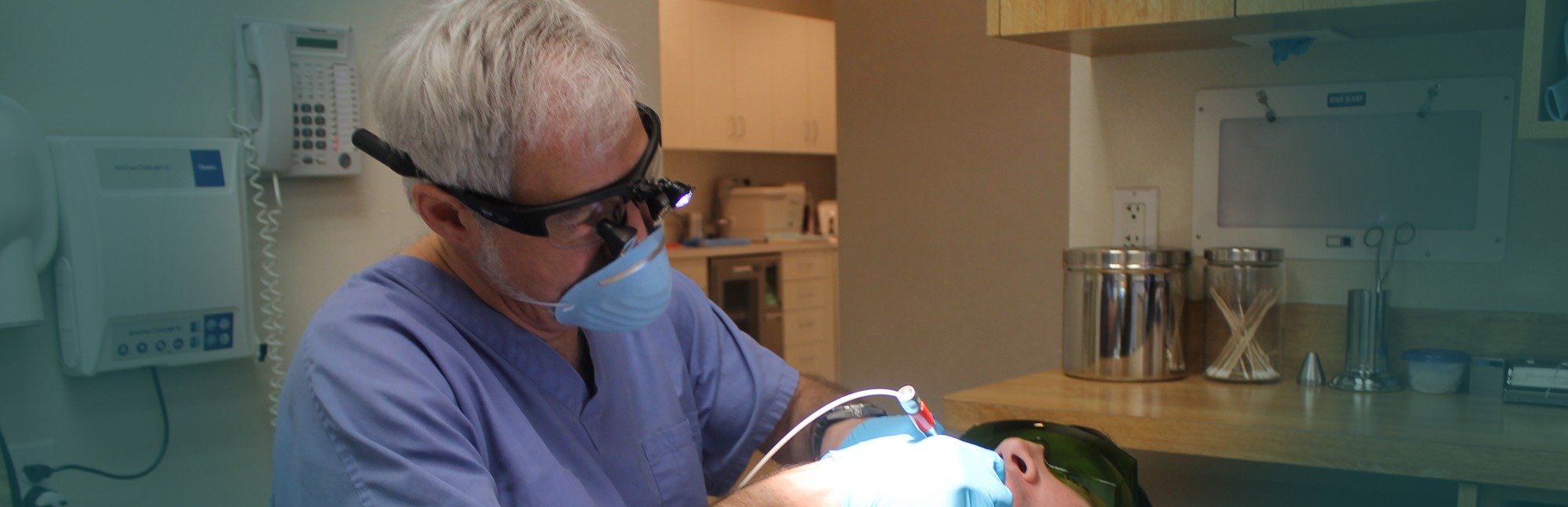
What we do to Assess for Oral Cancer
Vizilite
In our continuing efforts to provide the most advanced technology and highest standard of care available to our patients, this practice is proud to announce the inclusion of the ViziLite Plus exam as an integral part of our annual comprehensive oral screening program.
Why Should I get a ViziLite Screening?
One person dies every hour from oral cancer in the United States – and the mortality has remained unchanged for more than 40 years. Late detection of oral cancer is the primary cause that both incidence and mortality rates of oral cancer continue to increase.
As with most other cancers, age is the primary risk factor, and 27% of oral cancer victims have no lifestyle risk factors.
According to the American Cancer Society, more women in the United States will be diagnosed with oral cancer this year (12,000 cases) than will be diagnosed with cervical cancer (<10,000 cases), and there are as many cases of oral cancer caused by the human papilloma virus (HPV 16/18), a sexually transmitted disease, as there are HPV-related cases of cervical cancer.
Clinical studies have determined that using ViziLite Plus after the standard oral cancer examination improves the dental professional’s ability to identify and evaluate suspicious areas at the earliest stages. Early detection of pre-cancerous tissue can minimize or eliminate the potentially disfiguring effects of oral cancer and possibly save your life.
Proven screening technologies such as a mammogram, Pap smear, PSA testing and colonoscopy offer the same type of early detection of cancer. VizilLite Plus is an easy and painless examination that gives this practice the best chance to find any oral abnormalities you may have at the earliest possible stage.
Identafi® Oral Cancer Screening Device
Identafi® technology includes nineteen issued patents and seven patents pending relating to the detection of various types of pre-cancerous conditions (dysplasia) and cancers. The detection is made through the processing of optical fluorescence and reflectance in certain body tissues.
The Identafi® core technologies are based on high-speed, high-resolution capabilities from its patented optical processing technology and includes the ability to read metabolic and physiologic differences in diseased and healthy tissue in the human body. The core technology provides for the Identafi® technology platform development and introduction of cancer diagnostic products.
The screening product is housed in an easily portable unit designed for use in the offices of dentists, specialists, and general practitioners for screening and diagnosing cancers and pre-cancers. Products now in development will be able to detect pre-cancerous cells and cancers in specific areas of the body including, but not limited to, oral (the mouth, throat, tongue and tonsils), cervix, skin, bladder, esophagus and colon.
HOW IT WORKS
The deep penetrating power of the Identafi® multiple wavelengths will enhance the early detection of mucosal abnormalities that may be cancerous.
The new Identafi® uses the Identafi® Multi-Spectral Fluorescence and Reflectance technology to enhance visualization of mucosal abnormalities such as oral cancer or premalignant dysplasia that may not be apparent to the naked eye. But unlike other fluorescence technologies and dye systems, the Identafi® is Multi-Spectral with three distinct color wavelengths, making it easier to distinguish lesion morphology and vasculature aiding the dental professional in determining next steps.
The Identafi® uses white, violet and amber wavelengths of light to excite oral tissue in distinct and unique ways. As a result, biochemical changes can be monitored with fluorescence, while morphological changes can be monitored with reflectance.
The combined system of fluorescence and reflectance uses the body’s natural tissue properties as an adjunctive tool for oral mucosal examination. Conventional examination of tissue is performed using a highly concentrated Whitelight.
Wearing reusable Identafi® filtered eyewear to enhance visual effects and allow transmission of reflected light, the health professional then switches to Violet for a second observation.
The clinician’s photosensitive glasses block the violet excitation light and allow the observance of the tissues natural fluorescence. Violet light enhances normal tissue’s natural fluorescence; however, suspicious tissue appears dark because of its loss of fluorescence.
When suspect abnormalities are present the selector is switched to Amber light, which enhances normal tissue’s reflectance properties so the clinician may directly observe the difference between normal and abnormal tissue’s vasculature. This independent view of vascular architecture may assist in the assessment of confounders when screening for oral cancer.
Studies indicate abnormal tissue has a diffuse vasculature, where normal tissue vasculature is clearly defined. The combination of all three Multi-Spectral wavelengths provides the clinician with more visual information, improved results over direct visual exam alone and increased confidence for recommending biopsies.
VELscope®
 VELscope is a revolutionary hand-held device that provides dentists and hygienists with an easy-to-use adjunctive mucosal examination system for the early detection of abnormal tissue, namely oral cancer.
The VELscope handpiece emits a safe blue light into the oral cavity, which excites the tissue from the surface of the epithelium through to the basement membrane and beyond, causing normal tissue to fluoresce.
The clinician is then able to immediately view the different fluorescence responses to help differentiate between normal and abnormal tissue. In fact, VELscope is the only non-invasive adjunctive device clinically proven to help discover oral disease.
Discovering soft tissue abnormalities is particularly important in the fight against oral cancer. Because the VELscope® assists in early detection, cancer can be caught before it has time to spread, potentially saving lives through less invasive, more effective treatment.
Do Not Ignore Suspicious Lumps or Sores.
PATIENT INFORMATION
- Learn more about Dr. Goettishiem
- Schedule An Appointment Online
- New Patient Registration Forms
- Prepare for Your First Visit
- Dental Insurance Information
- Payment Options
- Get Driving Directions
Looking For Excellent Periodontal Care?
Dr. Goettisheim can place dental implants, treat gum disease, perform cosmetic periodontal surgery, and more. Contact us today!